
Introduction
Peripheral nerve entrapment can turn everyday movement into a persistent source of discomfort. Pressure, adhesions, scar tissue, inflammation, or restricted tissue mobility may affect a nerve as it passes through anatomically narrow spaces. Patients may experience pain, numbness, tingling, weakness, or reduced function, depending on the nerve involved.
For clinicians managing these conditions, the challenge is not simply identifying where pain occurs. The more important question is what is happening around the affected nerve and how precisely an intervention can reach the intended anatomical plane.
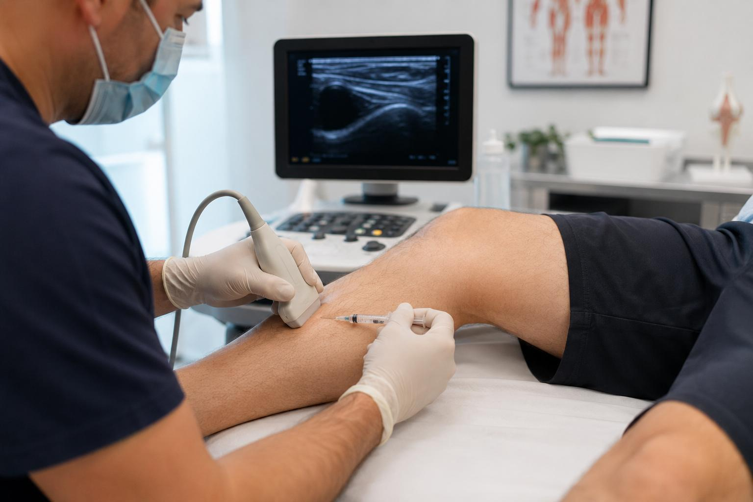
This is where ultrasound-guided nerve hydrodissection has attracted increasing clinical attention. The technique uses real-time ultrasound imaging to identify a peripheral nerve and guide the controlled injection of fluid around it. The injected solution can create separation between the nerve and surrounding tissues, potentially reducing mechanical restriction and improving nerve mobility.
For professional clinics, this development also highlights a broader trend in modern treatment technology: visualization is becoming as important as energy or treatment delivery itself. High-resolution ultrasound systems allow trained practitioners to see anatomical structures dynamically rather than relying only on surface landmarks or palpation.
For manufacturers and professional aesthetic-equipment providers, this distinction is important. Advanced imaging technology is not simply another treatment function. It can become part of a more precise clinical workflow in which assessment, visualization, intervention, and follow-up are connected.
Background/Problem: Why Peripheral Nerves Become Entrapped
Peripheral nerves travel through muscles, fascia, ligaments, tunnels, and other anatomical structures. In certain areas, a nerve may pass through a relatively narrow space where it becomes vulnerable to compression or irritation.
Common examples include the median nerve in carpal tunnel syndrome and the ulnar nerve around the elbow. Other peripheral nerves may also become symptomatic depending on anatomical structure, previous injury, repetitive movement, inflammation, or postoperative scar tissue.
The symptoms can vary considerably.
A patient may report:
- Localized or radiating pain
- Tingling or numbness
- Burning or electric-like sensations
- Muscle weakness
- Reduced grip or movement ability
- Sensitivity around a specific anatomical pathway
Traditional conservative management may include activity modification, splinting, physical therapy, medication, or other non-invasive approaches. When symptoms persist, clinicians may consider image-guided interventions or, in selected cases, surgery.
The difficulty is that peripheral nerves are relatively small structures. A procedure performed without adequate visualization may have less control over the exact anatomical target.
This is one reason ultrasound has become increasingly valuable in peripheral nerve assessment and intervention. Instead of treating the painful area as a general zone, ultrasound can help trained practitioners identify the nerve, surrounding fascia, vessels, tendons, and other relevant structures in real time.
From “Where Does It Hurt?” to “What Is Happening Around the Nerve?”
This change in perspective is important.
A patient may point to the wrist and say, “This is where it hurts.” A clinician, however, needs to understand whether the median nerve is enlarged, compressed, displaced, restricted by surrounding tissue, or affected by another anatomical factor.
The clinical value of ultrasound therefore extends beyond simply producing an image.
It provides anatomical information that can support clinical decision-making.
Technology/Mechanism: How Ultrasound-Guided Nerve Hydrodissection Works
Nerve hydrodissection is based on a relatively straightforward concept: using fluid to separate a peripheral nerve from adjacent structures.
The procedure begins with ultrasound assessment. The practitioner scans the target region and identifies the affected nerve along its anatomical course.
The ultrasound image can help evaluate features such as:
- Nerve location and continuity
- Cross-sectional appearance
- Surrounding fascia
- Tendons and muscles
- Blood vessels
- Potential areas of compression
- Postoperative or fibrotic tissue
Once the target anatomy has been identified, the practitioner can plan an appropriate needle path.
Under continuous ultrasound visualization, the needle is advanced toward the perineural tissue plane. Fluid is then introduced around the nerve in a controlled manner.
The objective is not to inject directly into the nerve.
Instead, the fluid is placed around the nerve to create a separation between the nerve and surrounding tissues. Depending on the clinical situation and injectate selected by the qualified practitioner, this may help reduce mechanical restriction and improve the nerve's ability to glide within its surrounding tissue environment.
Why Real-Time Visualization Matters
This is where the imaging system becomes central to the procedure.
Ultrasound provides a live view of the needle, nerve, surrounding soft tissues, and changing fluid plane. The operator can adjust the needle trajectory and observe tissue separation as the procedure progresses.
This is fundamentally different from relying only on anatomical landmarks.
For professional clinics, high-quality ultrasound equipment therefore provides value in three interconnected areas:
Visualization → Precision → Procedural Control
The first step is seeing the anatomy clearly. The second is identifying the intended tissue plane. The third is controlling the intervention according to what is visible on the screen.
This workflow explains why image quality, probe selection, depth control, contrast, resolution, and ergonomic system design matter in professional environments.
[IN-ARTICLE IMAGE — Recommended placement]
Suggested visual: A clean medical illustration showing a peripheral nerve before and after ultrasound-guided hydrodissection. The left side should show the nerve compressed by surrounding tissue; the right side should show the nerve surrounded by injected fluid and separated from adjacent structures. Add a small ultrasound probe and needle trajectory to visually explain real-time guidance.
What Can the Injected Fluid Do?
The term “hydrodissection” describes the mechanical separation produced by the injected fluid, while the biological effects may also depend on the selected injectate.
Different clinical protocols have investigated substances including saline, local anesthetics, corticosteroids, 5% dextrose in water, and platelet-rich plasma.
The choice should not be treated as a universal formula.
Different patients, nerves, clinical conditions, and treatment objectives may require different approaches. Current literature suggests that D5W and PRP have received particular research attention, while further investigation is still needed to determine the optimal injectate, volume, and patient selection for different nerve entrapment conditions.
This is another reason professional equipment and trained operators matter.
A sophisticated ultrasound system does not replace clinical expertise. Rather, it provides the visual information that allows qualified practitioners to apply their expertise with greater anatomical awareness.
Advantages/Comparison: Why Ultrasound Guidance Changes the Clinical Workflow
1. More Precise Anatomical Targeting
One of the most important advantages is direct visualization.
Instead of estimating the location of a small peripheral nerve from external landmarks, the practitioner can identify the structure on the screen and plan the intervention accordingly.
2. Real-Time Needle Visualization
Ultrasound can provide continuous visualization during needle advancement.
This allows the operator to monitor the relationship between the needle and nearby anatomical structures rather than treating the procedure as a blind injection.
3. Dynamic Assessment
Ultrasound is not limited to static images.
Soft tissues can be observed dynamically, and the operator can evaluate changes during scanning and intervention.
This makes ultrasound particularly relevant to professional practices where assessment and treatment need to be integrated into one workflow.
4. No Ionizing Radiation
Diagnostic ultrasound uses high-frequency sound waves rather than X-rays. This makes it an attractive imaging modality for many soft-tissue applications when appropriately used.
However, “non-ionizing” should not be interpreted as “risk-free.” Ultrasound equipment should be operated according to applicable regulations, manufacturer instructions, and professional safety principles.
5. More Information Than Surface Palpation
Palpation can provide useful clinical information, but it cannot directly show the nerve, vessels, fascial layers, or deeper soft-tissue relationships.
Ultrasound adds another layer of anatomical information.
Professional Ultrasound vs. Home Devices
The difference between professional and consumer-oriented equipment becomes particularly clear in this application.
A home-use device may focus on convenience, simple operation, or general wellness monitoring. A professional ultrasound system, by contrast, needs to support clinical workflows where image quality and anatomical interpretation are critical.
Professional users may prioritize:
- High-resolution imaging
- Appropriate transducer selection
- Adjustable depth and gain
- Clear tissue differentiation
- Real-time imaging
- Stable image processing
- Ergonomic probe handling
- Easy image review and documentation
- Reliable system performance
- Training and technical support
For medical and professional clinics, these features are not simply technical specifications. They influence how efficiently a practitioner can assess anatomy and make decisions.
For equipment manufacturers, therefore, the strongest product positioning is not necessarily “more power.”
It is better visualization, better control, and better integration into professional workflows.
Applications/Client Needs
Ultrasound-guided nerve hydrodissection has been studied most extensively in carpal tunnel syndrome, while selected reports and clinical discussions have also considered conditions involving the ulnar, radial, saphenous, sciatic, and fibular nerves.
However, suitability depends on individual diagnosis and clinical assessment.
Carpal Tunnel Syndrome
The median nerve passes through the carpal tunnel at the wrist. Compression in this confined anatomical space can produce numbness, tingling, pain, and weakness affecting the hand.
Because of its accessibility and relatively well-defined anatomy, the median nerve has become one of the most frequently studied targets for ultrasound-guided hydrodissection.
Ulnar Neuropathy
The ulnar nerve around the elbow is another potential target in selected cases.
Ultrasound can help practitioners follow the nerve dynamically and evaluate its relationship with surrounding anatomical structures.
Postoperative or Scar-Related Entrapment
Scar tissue can sometimes contribute to nerve restriction after surgery.
In appropriately selected cases, ultrasound may help identify the relationship between the nerve and surrounding scar tissue, supporting more precise procedural planning.
Professional Pain and Rehabilitation Clinics
The technology is particularly relevant to multidisciplinary environments that combine pain management, physical medicine, rehabilitation, sports medicine, or musculoskeletal assessment.
In these settings, ultrasound may become part of a broader clinical workflow rather than a standalone treatment device.
What Clinic Operators Should Look for in an Ultrasound System
For professional users, purchasing decisions should go beyond screen size or headline specifications.
A practical evaluation should consider whether the system can provide:
Clear peripheral nerve imaging: Small structures require adequate resolution and appropriate transducers.
Reliable real-time performance: Procedures involving needle visualization require stable imaging.
Flexible parameter adjustment: Depth, gain, focus, and other imaging controls can influence anatomical visibility.
Ergonomic operation: Long clinical sessions require comfortable probe handling and an efficient interface.
Image documentation: Saving and reviewing images can support clinical records and follow-up workflows where permitted.
Training and technical support: Advanced imaging technology is only useful when operators understand how to acquire and interpret appropriate images.
Regulatory suitability: Intended use, local regulations, device classification, operator qualifications, and facility requirements should all be evaluated before clinical deployment.
These factors are especially important for manufacturers supplying international professional markets. A device positioned for clinical imaging needs to be supported by appropriate technical documentation, quality systems, training materials, and regulatory strategy.
A Broader Trend: From Treatment Devices to Intelligent Clinical Platforms
The development of ultrasound-guided procedures illustrates a broader direction in professional equipment.
Modern clinical technology is increasingly moving from isolated treatment functions toward integrated workflows.
Instead of asking only:
“What energy does the device deliver?”
professional users increasingly ask:
“Can I see the target clearly?”
“Can I measure what I am seeing?”
“Can I control the procedure accurately?”
“Can I document the result?”
This shift has implications for the beauty and aesthetic-equipment industry as well.
Although nerve hydrodissection itself is a medical procedure and should not be marketed as a routine cosmetic treatment, the underlying technology trend is highly relevant: visualization, precision, and data-supported operation are becoming important differentiators for professional equipment.
For clinics that provide advanced aesthetic, rehabilitation, or medical services within their legal scope, imaging technology can contribute to a more structured approach to assessment and treatment planning.
Conclusion
Ultrasound-guided nerve hydrodissection represents a modern approach to selected peripheral nerve entrapment conditions. By combining high-resolution ultrasound imaging with controlled perineural fluid injection, trained clinicians can visualize the target anatomy and guide the intervention in real time.
Its value goes beyond the injection itself.
The real technological advantage lies in the combination of anatomical visualization, procedural precision, real-time feedback, and professional clinical control.
For equipment manufacturers and professional clinics, this technology also demonstrates an important direction for the future of medical and aesthetic equipment: advanced devices are increasingly expected to provide more than treatment output. They are expected to help professionals see, assess, control, and document.
At the same time, nerve hydrodissection remains a specialized clinical procedure. It should be performed only by appropriately trained and authorized healthcare professionals using equipment suitable for the intended clinical application. It should not be confused with consumer beauty treatments or routine home-use devices.
As ultrasound technology continues to improve, high-quality imaging platforms may play an increasingly important role in professional clinical environments where precision and anatomical understanding are essential
Founded in 2008, Guangzhou GLM Beauty Spa Equipment Factory specializes in research, development, manufacture and selling of beauty and slimming machines. Our production base is located in Guangzhou City, Guangdong Province, China. Since our foundation, we have introduced advanced technologies and equipment, to assure high quality and high performance of our products. With professional marketing guide and excellent after-sales service, our products have been exported to over 20 countries and regions, including Southeast Asia, the Middle East, Europe and the United States, won worldwide praises and gained a good reputation